
Josef Feit
Hypertext atlas of dermatopathology is meant for students of pathology, dermatology and dermatopathology. It contains clinical and histological images of skin diseases together with some introductory texts.
Hypertext atlas of pathology is meant for pregraduate students of pathology and image resource for teachers. It contains clinical, autoptic, endoscopic, CT, MRI, angiographic and histological images.
Both atlases offer histological images of high quality. Recently virtual microscope interface is used to access the high resolution images.
Images are annotated, so that arrows pointing to the important parts of an image can be activated.
Atlas is available on www.muni.cz/atlases
MSIE and Mozilla family of browsers are supported. The browser must have JavaScript enabled.
Each image in the atlas is linked with the reference to the contributor of the specific case. Reference pages sorted according to contributors are available.
After clicking on the link the window with the image in basic size will open. The list of signs can be activated and arrows will appear when user moves the mouse over the list.
Multilevel images (namely from CT or MRI scanners) can be controlled by arrows in the upper part of the image window.
Window with high resolution image will open after clicking on the button with magnifier. Its contens will be sychnronized with clicking in the basic size window.You can have any number of picture windows open at the same time. Each window is provided with a row of buttons (but not all of them are always available):
 opens or closes the window with the list of signs
 opens the magnifying window
 closes the magnifying window
 closes the window with pictures
 focusing up, changing the examination level (CT, MRI)
 focusing down, changing the examination level (CT, MRI)
 enlarging the basic picture, which can be dragged
 and similar buttons control the color planes of FISH images
This is what you should see while running this atlas: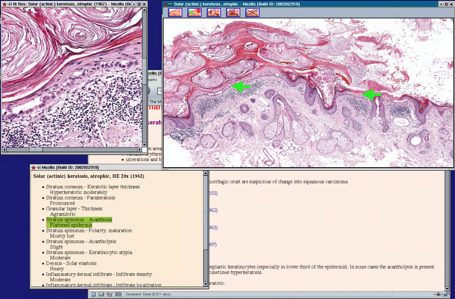
In the background there is the window with
the atlas itself. The main picture window
(with arrows activated) is in the foreground, right.
To the upper left there is a small window
showing the detail (the detail is driven
by mouse clicking in any site of the picture
in the main window, see bellow).
To the lower left there is a window with
the list of signs; each sign can activate
corresponding arrows in the main picture
window.
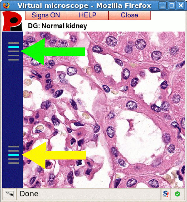
The latest improvement of the atlas is the virtual microscope. A small window with image detail will be open and synchronized with clicking into the basic window. In addition it will be possible:
Only the necessary parts of the image will be downloaded, saving time and memory of your computer.
Pictures
Macroscopic image with possible enlargement, rectal carcinoma infiltrating into the vagina:
 Adenocarcinoma of the rectum spreading into vaginal wall, Macro (71888)
Histological image: infiltrating carcinoma of the stomach (classic approach to the detail), HE, 40×:
Infiltrating carcinoma, stomach, HE 40x (70008)
Histological image, focusable: mycosis of the esophagus, PAS, 40×:
Candidosis of the esophagus, PAS 40x (70034)
Histological image, FISH: follicular lymphoma, t(14;18)(q32.3;q22), 40×:
Follicular lymphoma, secondary skin infiltration in nodal lymphoma, FISH 40x (71887)
Histological image, virtual microscope, cortex of normal kidney, 100×:
Normal kindey, cortex, HE 100x (72474)
Histological image, fullscreen zoomify (www.zoomify.com), meconial changes, placenta 40×:
Placenta, abortion; surface reaction to meconium, HE 40x (71958) [zoomify]
Scanscope 2 histological image, fullscreen zoomify (www.zoomify.com), burned out lichen planus, 20×:
Lichen planus atrophic, HE 20x (4436) [zoomify]
CT scan, multiple levels: adenocarcinoma of the rectum with destruction of sacral bones:
Adenocarcinoma of the rectum, osteolysis of the sacral bone, CT, X-ray (71828)